Therapeutic Cloning: Hope or Hype?
by Janet D. Rowley
 here is no one that I am aware of on the President's Council on Bioethics who would be supportive of cloning a human being. Apart from the many ethical issues, there are medical and scientific reasons to support not cloning a human being--or as the council's chairman, Leon Kass, calls it, "baby making"--at this time. Here, I will explain the differences between human reproductive cloning and therapeutic stem cell cloning, and will review the risks and possibilities of each.
How human reproductive cloning works
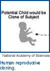
 In order to clone a human, you have to have a woman willing to provide eggs. She would then be treated with hormones in order to release many eggs, not just a single one, and the eggs would need to be surgically removed. There is certainly some risk in the procedure to the egg donor, and while it is not a major risk, at the present time this course of action would have to be done with the woman's informed consent. After the eggs are removed from the donor, the nuclei of the eggs (where the DNA is stored) are taken out to create the "Enucleated Egg" as shown in Figure 1. The rest of the cellular material is cytoplasma, which provides nutrients for the eggs' survival.
Once you remove the DNA from the donor's eggs, you then obtain a cell from the patient or subject. Various tissues could be used, and one method is to take a little snip of skin and culture it in a petri dish. This gives you multiple cells from this patient. What you really want is the nucleus--the DNA of that patient. You then take the nucleus out of the cell, put it in a glass tube, and insert the glass tube into the egg. Then, with a little bit of force, you place the nucleus into the egg (see "Nucleus Transfer" in Figure 1). The result is that you now have an egg with DNA and the DNA comes from the patient or the subject. You then must activate the nucleus, or trick it into thinking the ovum has been fertilized, so that the cell begins to divide.
When the cells divide, such as into eighth and sixteenth cell stages (see "Morula", Figure 1), very frequently--and this is done in in vitro fertilization--at least one cell is removed to perform gene analysis. If you are trying to screen the ovum for the presence of a genetic defect that is inherited in the family, then you remove a single cell, leaving the other cells undisturbed so they will grow normally. You then complete your DNA testing on this one cell, looking for whatever genetic defect for which you are testing.
The remaining cells then divide and form the blastocyst--a cluster of cells created during the 64-200 cell stage that will grow to be the embryo (see "Blastocyst" in Figure 1). The rest of the cells around the outside will form the placenta. If you were going to attempt reproductive cloning, at this stage you would then implant this blastocyst into the woman's uterus. She would be prepared with hormones so her uterus would be receptive to the blastocyst and, if all goes well, she would have a developing fetus and finally give birth successfully to a child.
Risks in human reproductive cloningOne of the risks, aside from moral issues, associated with reproductive cloning is that for reasons we do not entirely understand, very often (and this data comes from experimental animals) the fetus gets rather large and can actually cause potential difficulty at the time of birth.
Another risk is that reproductive cloning is not a highly successful procedure. In a 2002 report from the University of Pennsylvania Veterinary School, scientists have now have identified a gene, Oct4, which is active in the oocyte and sperm, but inactive in adult cells. One of the things that has to happen to have a successful transition to each stage in the blastocyst is to have the Oct4gene, which is off and not expressed, inactive in this somatic (or body) cell. It somehow has to be turned on due to signals from cytoplasma of the ovum to allow this kind of development.
What turns on or expresses Oct4 in this particular circumstance is not clear at the present time, but there are probably a series of signals (from regulatory genes) that enable this to happen. At present, we do not know what else is operating and we do not know how to automatically turn on Oct4. To make science work, once you understand the problem, you have to figure out how to do it. You focus on that gene and the regulation for that particular gene as well as collaborating genes.
Inefficiencies in animal reproductive cloning
The National Academy of Sciences reported on the success rate of cloned embryos created in experimental animals. In one report, there are well over 1,000 embryos created, but which result in very few live births; essentially 1 percent, in many of these instances, result in live births. There were experiments in Oregon on monkeys some time ago, but the results had been so discouraging that this particular project has been stopped at least for now. If you translate data from monkeys to people, you see that the likelihood that you really are going to be successful with cloning a human child, given present technology, is extremely small.
Stem cell cloning
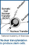
 You will recognize that Figure 2 is very similar to Figure 1. However, Figure 2 charts the creation of stem cells that can be used for various strategies and disease treatments.
Like in Figure 1, a woman must provide several eggs. The DNA is then removed from the ova and the nuclei from the patient or subject providing the somatic cells is likewise transferred. Again, using in vitro fertilization, this is done in a lab environment. In Figure 2, you will see an enlargement of the "Blastocyst" stage that shows that the inner cell mass gives rise to the embryo and the "trophoblast" gives rise to the placenta.
In stem cell cloning, you then remove these cells, grow them in tissue culture, and create the pluripotent embryonic stem cells with which scientists will be working. There are several things to point out here. One is that these cells (see "Pluripotent Embryonic Stem Cells" in Figure 2) in fact are growing on a layer of other cells; they are not just sitting there in a glass or a plastic petri dish. A layer of fiberglass allows other cells at the bottom to attach to the petri dish; our cells are growing on top of them. This "feeder layer" provides growth factors, many that we do not understand at the present time, but it is clear that they are very important. For all of the embryonic stem cell lines that have been derived so far, this feeder layer is known to have existed. The FDA is in fact treating these cells as xenografts, because the human cells have been grown in contact with animal cells and the possibility of virus transmission cannot be excluded in this situation.
Next, you take the cells now growing in tissue cultures, separate them, and put them into specialized environments with growth factors in feeder layers that will encourage these pluripotent stem cells. These can grow into different specialized cells, such as heart muscle cells, liver cells, or bone marrow cells (which would help in bone marrow transplants). At present, there is a great deal of emphasis placed on pancreatic islet cells, which could be transplanted into an individual with diabetes. One hope is that for a diabetic individual, cells from the individual could be grown in this manner and then given back to this individual to help treat the diabetes. Because the DNA in the nucleus is actually derived from the individual, you would not have some of the problems with immune rejection or transplantation rejection. One of the things that cannot be ignored, however, is that there are also special kinds of DNA in the cytoplasma of the ovum which would be foreign to this individual and therefore could elicit some immune response. However, this response is not as strong an immune response as you would get with foreign DNA.
The other point that I want to make is that you have to make sure that you get very pure specialized cells that you will be injecting into experimental animals. If you inject pluripotent stem cells into an experimental animal, it will result in tumors, so that is an additional caveat or concern.
Other means of obtaining human stem cellsThere are other ways to obtain human stem cells. The strategy that has been used by Dr. Gearhart at Johns Hopkins University is to use therapeutically aborted fetuses (these fetuses have reached a certain stage of development, around 15 weeks) as the region of the fetus that will encase the primordial germ cells for that fetus were it to grow sperm and be an adult. Dr. Gearhart would use those germ cells from the fetus as the source for his strategy.
Individuals such as James Compton in Madison, Wisconsin, as well as Dr. Gearhart and other scientists around the world, have actually developed cell lines out of these so that you end up with embryonic stem cells that you can actually grow in tissue cultures.
In August 2002 President Bush said that he would permit and support research using 60 embryonic stem cell lines that have already been derived. There are a number of problems with this. Firstly, many of these lines have not been studied with sufficient care to know that in fact they are a permanent cell line. Some of them were already growing for a short period of time. Another important concern is that cells in tissue cultures often undergo mutations; the DNA may have mutations or obvious chromosome abnormalities, such as an extra chromosome, a missing chromosome, or translocations. Most of those cell lines have not been analyzed carefully to determine whether they have 46 normal chromosomes and that they appear to be normal.
There are a number of individuals who believe that scientists do not need to use embryonic stem cells, but rather adult stem cells. Catherine Verfaillie at the University of Minnesota has discussed this work in public, arguing that you could take adult stem cells and then use the same procedures to get the adult stem cells in cultures to develop some of the specialized cells you need. There are several problems, however. There is no evidence that the adult stem cells can develop into all of the specialized cells that embryonic stem cells can. Some specialized cells have developed; Verfaillie has developed what she believes are nerve cells and muscle cells, but has not yet derived blood cells. The other thing that we know is that, at least in nature, these cells still have a limited time to divide, so that after 60-70 generations an animal cell can no longer divide. This is assumed to be associated with the fact that the ends of the chromosomes get shorter with each cell division, much like a biological clock telling the cell it cannot divide any more. A cell developed from adults, of course, would already have used up some proportion of their lifetime, if you will, whereas the embryonic stem cells would have a much, much longer time. These are some of the issues.
RecommendationsI have not touched on any of the ethical issues that individuals have raised concerning human reproductive and therapeutic stem cell cloning. I do want to say that in its report the National Academy of Sciences provides a number of recommendations, and one of the most important is that therapeutic cloning should be allowed. Reproductive cloning, however, should be banned and should carry criminal penalties to force people to pay attention and not break the law.
The other recommendation is to establish a national advisory or regulatory board broadly constituted that would review all scientific proposals for both scientific merit and as well as making certain that new procedures do not pose undue risks to the individuals involved. This has precedence. In the early 1970s when scientists realized that they could take a piece of DNA from one organism and transfer that DNA into another organism, there were great risks, but the risks at that time were not well known. At that time, scientists advocated we set up a review committee that makes sure that these experiments are done in the appropriate setting to minimize the risk of release of these foreign DNAs, and to ensure the experiments have some scientific merit to them.
Relevant linkThe President's Council on Bioethics
(www.bioethics.gov)
ABOUT THE AUTHOR |
Janet D. Rowley
Janet D. Rowley, M.D., D.Sc., is Blum-Riese Distinguished Service Professor of Medicine, Molecular Genetics and Cell Biology, and Human Genetics at the Pritzker School of Medicine at the University of Chicago. Dr. Rowley is internationally renowned for her studies of chromosome abnormalities in human leukemia and lymphoma. She is the recipient of the National Medal of Science (1999) and the Albert Lasker Clinical Medicine Research Prize (1998), the most distinguished American honor for clinical medical research.
COPYRIGHT | The text of this article is drawn from a talk given on May 15, 2002, at the Franke Institute for the Humanities at the University of Chicago. Copyright 2003 The University of Chicago.